Diffusion Tensor Imaging Traumatic Brain Injury
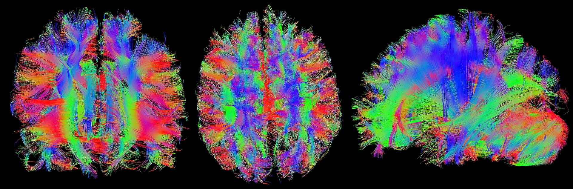
Diffusion Tensor Imaging (DTI) has become increasingly common in traumatic brain injury lawsuits as a means to argue the presence of brain damage that does not appear on any other forms of neuroimaging. DTI is an experimental MRI technique that measures how water molecules flow in the brain. Often plaintiff experts claim that DTI can detect changes in white matter (nerve fibers between neurons in the brain) that are undetectable by other forms of imaging, and cite this as evidence of brain damage. However, more and more research shows that DTI detects white matter changes from causes other than trauma. For instance, a study this month out of the University of Alberta examined young adults between the ages of 14 to 17 with a history of mental health issues (depression, anxiety, ADHD) to determine the functioning of their white matter. Each individual received DTIs to examine the white matter within their brain. These scans were then compared to a second set of adolescents of the same age group who had no history of mental illness.
The conclusion of the study showed a clinically significant difference in the connections of the neural pathways in those previously diagnosed with a mental health issue. Compared to their healthy peers, those with mental health issues had more difficulty in their decision-making and cognitive control. In fact, the results tell us these adolescents have “less neural efficiency” which has impacted not only their attention in general, but their ability to focus across their different environments.
This study again shows that differences on DTI are not per se evidence of a TBI. It is essential in any case involving DTI to make sure to understand not only the imaging findings and their implications for a particular individual, but also to explore the specific reliability and methodological shortcomings inherent to this form of neuroimaging.